Ready to Schedule?
Save time by selecting your own appointment slot through our secure scheduling portal.


Medically reviewed by Miller Vein
Ever notice small red or blue veins on your legs?
Or maybe you’ve seen thicker, bulging veins that twist beneath the skin?
Find out how we treat spider veins before your visit.
While these visible veins can be confusing or a little alarming, understanding what they are can help you take the right steps to keep your legs healthy and beautiful.
So, when it comes to spider veins and varicose veins, what’s really the difference?
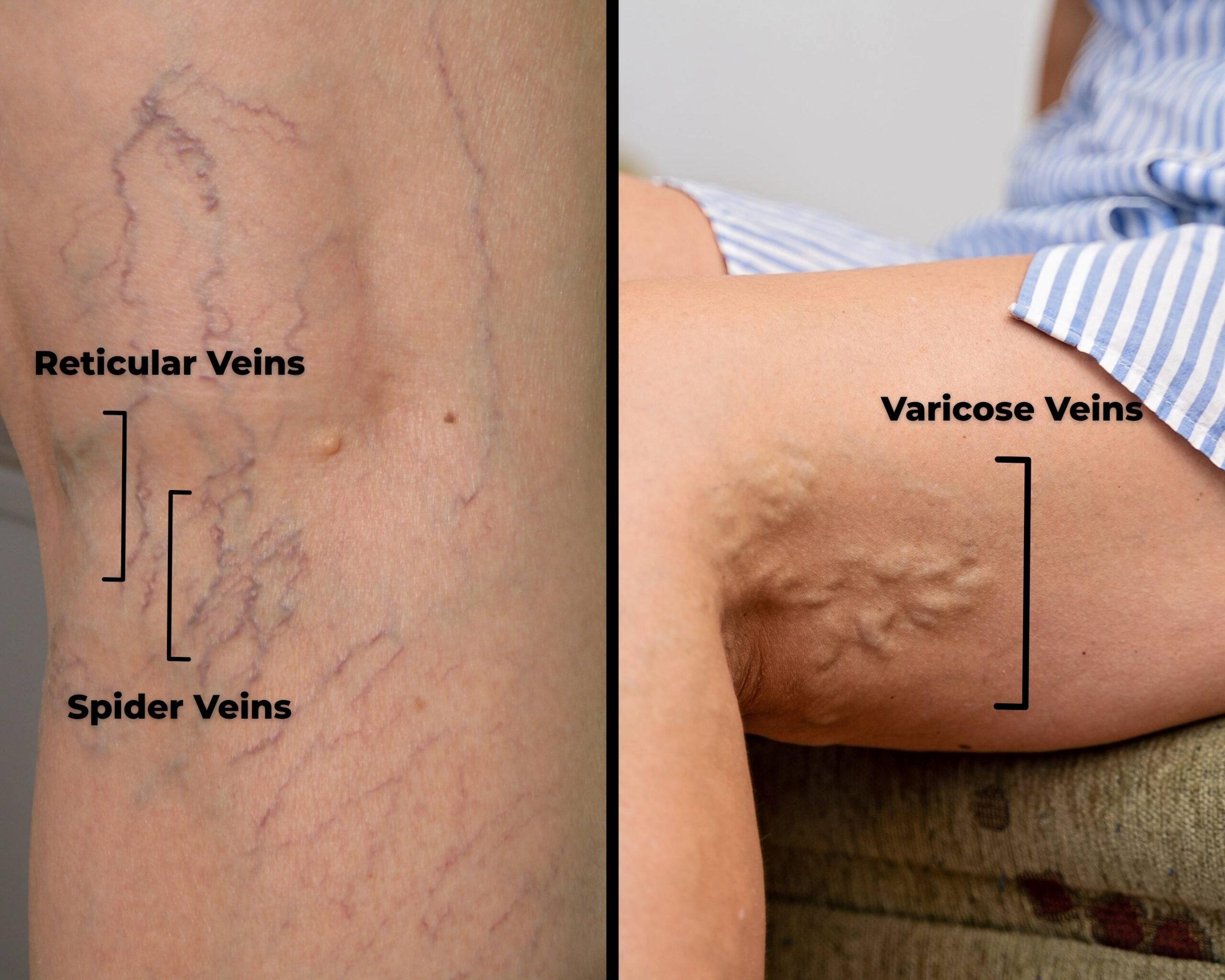
Spider veins are the tiny red, purple, or blue veins that often appear on the thighs, calves, or even the face. They’re called “spider veins” because they can look like little spider webs.
Although spider veins are usually a cosmetic issue, they can sometimes point to deeper vein problems. They may also cause itching or mild discomfort.
One study revealed that 50% of people with spider veins had symptoms. Out of 200 patients treated with sclerotherapy, 53% had symptoms (such as tiredness, pain, or burning), and 85% reported relief following treatment. (Weiss and Weiss, J Derm Surg Oncol, 1990)
If you notice a cluster of spider veins around your ankle, it could be a sign of a more serious condition called chronic venous insufficiency (CVI). This condition often comes with symptoms like leg heaviness, aching, swelling, throbbing, or itching.
Reticular veins are the “middle child” of visible veins. They’re larger than spider veins but smaller than varicose veins.
These bluish-green veins lie just under the skin and may not bulge, but they’re often visible on the back of the legs or behind the knees.
Varicose veins are the ones that get the most attention. That’s for good reason. These thick, rope-like veins bulge under the skin, often appearing twisted and swollen.
Unlike spider veins, varicose veins are more than a cosmetic issue. They are part of a medical condition known as venous reflux disease or, when more severe, chronic venous insufficiency. These veins cause significant symptoms, and therefore treatment is covered by insurance.
Understanding the difference between spider veins vs. varicose veins can help you take action early—whether that means improving circulation with lifestyle changes, wearing compression stockings, or seeking treatment.
In-office procedures today are minimally invasive, highly effective, and require no downtime.
And remember: what you see on the skin is only part of the picture. A vein specialist can use ultrasound to look deeper and guide the best treatment.
1. Caggiati A, et al. Nomenclature of the veins of the lower limbs: An international interdisciplinary consensus statement. Phlebology. 2005.
2. Labropoulos N, et al. Classification of venous disease: Guidelines and practical applications. J Vasc Surg. 2004.
3. Eberhardt RT, Raffetto JD. Chronic venous insufficiency. Circulation. 2014;130(4):333–346.
4. Parsi K. Reticular veins: Definitions and management. Australasian Journal of Dermatology. 2017;58(2):87–91.
5. Gloviczki P, et al. The care of patients with varicose veins and associated chronic venous diseases: Clinical practice guidelines. J Vasc Surg. 2011;53(5 Suppl):2S–48S.
6. Weiss RA, Weiss MA. Treatment of reticular and spider veins with sclerotherapy and lasers. Dermatol Surg. 2000;26(5):403–414.
Prefer a call? Fill out the form below, and our team will reach out to help you coordinate your visit.
"*" indicates required fields
Save time by selecting your own appointment slot through our secure scheduling portal.
